A Nuclear medicine scan is a diagnostic test that uses a small amount of radioactive material to show how your organs and tissues are functioning, not just what they look like. Artesia General Hospital (AGH) offers nuclear medicine imaging at our Artesia, NM campus using the GE HealthCare NM 830 gamma camera, serving patients from Artesia, Carlsbad, Roswell, Hobbs, and across southeastern New Mexico.
You may also hear this test called a nuclear scan, nuclear imaging, radionuclide imaging, or SPECT scan. These terms all refer to the same type of procedure. If your physician ordered a “nuclear medicine” study or a “SPECT” scan, you are in the right place.
If the name sounds alarming, that is understandable. Words like “radioactive” and “nuclear” carry associations that have nothing to do with what actually happens in the imaging department. A nuclear medicine scan is safe, painless, and non-invasive. The radioactive material used is a very small, carefully measured amount that leaves your body within hours. And the diagnostic information it produces gives your physician a picture of organ function that no other imaging test can provide.
This guide explains what nuclear medicine imaging is, which conditions it helps diagnose, what the GE NM 830 system means for your experience as a patient, and exactly what to expect from your appointment at AGH.
What Is Nuclear Medicine Imaging?
Most diagnostic imaging tests — X-rays, CT scans, MRIs — produce pictures of physical structures: the size, shape, and position of bones and organs. Nuclear medicine imaging adds a fundamentally different layer of information: it shows how those structures are working at the cellular level.
The test works by introducing a small amount of radioactive material called a tracer, or radiopharmaceutical, into your body. The tracer is designed to travel to a specific organ or tissue. Once there, it emits tiny amounts of energy in the form of gamma rays. The GE NM 830’s dual-head gamma camera detects those gamma rays and converts them into detailed images that your radiologist interprets.
According to the Society of Nuclear Medicine and Molecular Imaging (SNMMI), nuclear medicine procedures can detect disease in its earliest stages, often before it would appear on a CT scan or MRI. This makes it especially valuable for evaluating heart disease, cancer, thyroid disorders, bone conditions, and kidney function.
The GE HealthCare NM 830: What It Means for You
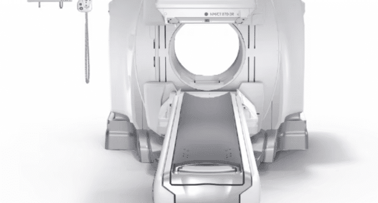
AGH recently installed the GE HealthCare NM 830, a current-generation dual-head SPECT gamma camera. The NM 830 uses two detector heads positioned around the patient, capturing imaging data from multiple angles simultaneously for more comprehensive SPECT images in a single pass.
For patients, the most meaningful improvements over older equipment are:
- Shorter scan times: Evolution technology reduces scan time or injected tracer dose by up to 25 percent compared to prior-generation software, meaning less time lying still and a more comfortable experience.
- Open design: The NM 830’s Elite NXT detectors are 13 centimeters slimmer than the previous generation. The detectors do not enclose you — you lie on an open table while the camera moves around you. Most patients find this system far less confining than they expected.
- Improved image quality: Reduced analog noise and high-resolution collimators produce sharper images, giving your radiologist greater diagnostic clarity.
Types of Nuclear Medicine Scans Available at AGH
The specific scan your physician orders depends on which organ or condition needs evaluation. Common nuclear medicine studies performed at AGH include:
| Scan Type | What It Evaluates | Common Reasons Ordered |
| Bone Scan | Bone structure, activity, and density | Unexplained bone pain, fracture evaluation, arthritis, cancer staging |
| Cardiac Scan / Nuclear Stress Test | Blood flow to the heart muscle at rest and under stress | Chest pain, suspected coronary artery disease, post-heart attack evaluation |
| Thyroid Scan | Thyroid gland structure and function | Thyroid nodules, hyperthyroidism, hypothyroidism, thyroid cancer follow-up |
| Renal (Kidney) Scan | Kidney blood flow, drainage, and function | Kidney obstruction, high blood pressure evaluation, post-transplant monitoring |
| Gallbladder (HIDA) Scan | Gallbladder function and bile flow | Abdominal pain, suspected gallbladder disease, post-surgery evaluation |
| Lung (V/Q) Scan | Air and blood flow in the lungs | Suspected pulmonary embolism (blood clot in the lung) |
| Gastric Emptying Scan | Speed of stomach emptying | Nausea, vomiting, abdominal pain after eating, suspected gastroparesis |
| Parathyroid (Sestamibi) Scan | Parathyroid glands function and localization | Elevated blood calcium levels, abnormal PTH levels |
If you are unsure which scan your physician has ordered, the AGH imaging scheduling team can clarify before your appointment. To schedule, call 1-855-RAD-EXAM (1-855-723-3926).
How Nuclear Medicine Differs From CT and MRI
A CT scan uses X-rays to produce detailed cross-sectional images of anatomy: structure, size, and position. An MRI uses magnetic fields and radio waves to produce high-contrast images of soft tissue. Both show what something looks like. Nuclear medicine shows how something is working.
As an example: a CT scan of the heart can show the anatomy of the coronary arteries. A nuclear stress test can show whether blood is actually flowing normally through those arteries when the heart is under stress. Both types of information are clinically valuable, and physicians often order them together. For some conditions, nuclear medicine provides diagnostic information that simply cannot be obtained any other way.
Is a Nuclear Medicine Scan Safe?
Yes. The radioactive tracers used in nuclear medicine are administered in very small, carefully measured amounts, individually calibrated for each patient and each exam type. According to RadiologyInfo.org, published by the Radiological Society of North America and the American College of Radiology, there are no known long-term adverse effects from the low-dose radiation exposure involved in nuclear medicine diagnostic procedures. Physicians have been using nuclear medicine diagnostic procedures for more than six decades.
The NM 830’s Evolution technology can reduce the injected tracer dose by up to 25 percent compared to prior-generation protocols while maintaining image quality, which is a meaningful improvement in the radiation profile of the exam.
Side effects are rare and usually mild. The most common is minor discomfort or a brief cold sensation moving up the arm when the tracer is injected intravenously, similar to any IV placement. Allergic reactions to radiotracers are extremely rare. If you have had a reaction to a radiotracer in the past, tell the scheduling team when you book your appointment.
Tell your physician and the AGH imaging team before your scan if you are pregnant, breastfeeding, or think you might be pregnant. See the FAQ section below for more detail on both situations.
What to Expect: Step by Step
Before Your Scan
Preparation varies by scan type. The AGH imaging team will give you specific instructions when you schedule. In general:
- Wear comfortable, loose-fitting clothing without metal fasteners, zippers, or underwire.
- Leave jewelry and valuables at home.
- Fasting is required for some scans, particularly cardiac and gastric emptying studies. Your scheduling team will confirm whether this applies to your exam.
- Tell the scheduling team about all medications you take. Some medications need to be held before certain scans, particularly cardiac stress tests.
- Arrive on time. The radioactive tracer is prepared specifically for your appointment time. Late arrival affects tracer quality and image results.
- Tell the team if you are pregnant, breastfeeding, claustrophobic, or have allergies or prior reactions to radiotracers.
When You Arrive
You will check in at the AGH imaging department and complete any necessary paperwork. A licensed nuclear medicine technologist will review your physician’s orders, confirm your identity, and walk you through the procedure before it begins. This is a good time to ask any questions you have.
Receiving the Tracer
For most scans, the tracer is administered through a small IV line placed in your arm. The injection takes only a few seconds. You may feel a brief sting when the line is placed and occasionally a cold sensation moving up the arm as the tracer is injected — both are normal and pass quickly. For thyroid scans, the tracer is taken orally as a capsule. For lung scans, part of the tracer may be inhaled.
After administration, there is often a waiting period before imaging begins, allowing the tracer to travel to the target organ. Waiting times vary: from about 30 minutes for some cardiac studies to two to four hours for bone scans. For bone scans specifically, you may be able to leave the facility during the waiting period and return for imaging — ask your technologist when you arrive. Bring something to read or listen to if you are staying on-site.
During the Scan
You will lie on a padded imaging table. The NM 830’s two detector heads will be positioned around you and move slowly during the scan. They do not enclose you — the open table design means you have clear sightlines throughout the procedure. The system is quiet and does not produce the loud sounds associated with MRI.
The most important thing is to remain still. Movement blurs the images. The imaging portion of most scans takes 20 to 45 minutes. Your technologist will stay nearby and can communicate with you throughout.
After Your Scan
When imaging is complete, the technologist removes the IV line if one was used. You can typically resume normal activities immediately unless your physician has instructed otherwise.
Drink extra water after your scan to help flush the tracer from your body more quickly. When using the restroom, put the toilet lid down and flush immediately after — this is a routine precaution to minimize contact with any tracer that leaves through urine.
Your images will be reviewed by a board-certified radiologist with nuclear medicine training. A report goes to your ordering physician, typically within one to two business days. Results go to your physician first — they will contact you to discuss findings and next steps. If you have not heard back after a few days, contact your ordering physician’s office directly.
Why Choose AGH for Nuclear Medicine in Southeastern New Mexico
For patients in Artesia, Carlsbad, Roswell, Hobbs, and across Eddy County, Chaves County, and Lea County, AGH means you do not have to travel to Albuquerque, El Paso, or Lubbock for quality nuclear medicine imaging. AGH is a DNV-accredited, nonprofit community hospital that has served southeastern New Mexico since 1939, with a full-time imaging team that includes licensed nuclear medicine technologists and board-certified radiologists.
The GE NM 830 gives patients in this region access to current-generation SPECT technology at a community hospital close to home — the same diagnostic capability available at larger systems, without the drive.
To schedule, a physician’s referral is required. Call 1-855-RAD-EXAM (1-855-723-3926) after receiving your order.
Frequently Asked Questions
What is nuclear medicine imaging used for?
Nuclear medicine imaging evaluates how organs and tissues are functioning, not just what they look like. Common uses include diagnosing heart disease, evaluating thyroid disorders, detecting cancer spread to bones, assessing kidney function, identifying gallbladder problems, and evaluating unexplained bone pain.
What are other names for a nuclear medicine scan?
Your physician may have used one of several terms: nuclear scan, nuclear imaging, radionuclide imaging, SPECT scan, or gamma camera scan. These all refer to the same general type of procedure. The specific name of your exam, such as bone scan, cardiac stress test, or thyroid scan, refers to which organ is being evaluated.
Is nuclear medicine safe?
Yes. Tracers are administered in very small, carefully measured amounts. According to RadiologyInfo.org, there are no known long-term adverse effects from the low-dose radiation exposure involved in nuclear medicine diagnostic procedures — physicians have used them safely for more than six decades. Side effects are rare and usually mild, most commonly minor discomfort at the injection site or a brief cold sensation in the arm.
Will I be radioactive after my scan?
Technically yes, but in a very small amount that poses no risk to others. Most tracers used in nuclear medicine clear the body within 24 to 48 hours, primarily through urine. You do not need to isolate yourself or avoid contact with others after most nuclear medicine scans. Drinking extra water speeds up the clearance process. Your technologist will give you any specific precautions relevant to your exam type.
Are there side effects?
Side effects are rare. The most common is minor discomfort when the IV line is placed, and occasionally a brief cold sensation moving up the arm as the tracer is injected — both are normal and pass within seconds. Allergic reactions to radiotracers are extremely rare. If you have had a prior reaction to a radiotracer, tell your scheduling team before your appointment.
What if I am claustrophobic?
The GE NM 830 at AGH uses an open table design — the detectors do not enclose you, and you have open sightlines throughout the scan. Most patients who expected to feel confined find the procedure much more tolerable than anticipated. If you have significant claustrophobia, tell your physician before your appointment. They may be able to prescribe a mild anti-anxiety medication to take beforehand. If you do take medication for claustrophobia, arrange for someone to drive you home.
I am breastfeeding. What do I need to know?
Some nuclear medicine tracers can pass into breast milk in small amounts. Depending on the type of scan ordered, you may need to pause breastfeeding for a period after your scan — from a few hours to longer for certain tracer types. Your nuclear medicine technologist will give you specific guidance based on your exam. As a general precaution, consider pumping and storing milk before your appointment so you have it available during any required pause. Discuss the timing with your technologist when you schedule.
Can I leave between my injection and my scan?
For scans with long waiting periods, such as bone scans, yes — you can typically leave the facility after the tracer injection and return when it is time to image. Your technologist will confirm whether this applies to your specific exam and give you a return time. If you leave, continue drinking water and avoid vigorous exercise during the wait.
How long does a nuclear medicine appointment take?
Total appointment time depends on the scan type. Some nuclear medicine studies take under two hours from arrival to completion. Bone scans typically require a two to four hour wait between tracer injection and imaging, so the full appointment can run three to five hours including imaging time. Your scheduling team will give you a full time estimate when you book.
How and when will I get my results?
Your images are reviewed by a board-certified radiologist and a report is sent to your ordering physician, typically within one to two business days. Results go to your physician first — they will contact you to discuss findings and recommend next steps. If you have not heard back within a few days of your expected results window, call your ordering physician’s office directly.
Are there any precautions after the scan?
For most standard nuclear medicine scans, precautions are minimal. Drink extra water to help flush the tracer. When using the restroom, put the toilet lid down and flush immediately as a routine precaution. You can resume normal activities right away unless your physician has given specific instructions. If you took anti-anxiety medication for claustrophobia, arrange a ride home and avoid driving for the rest of the day.
Can I drive myself home?
Yes, for most nuclear medicine scans. The exception is if you took medication for claustrophobia before your appointment — in that case, arrange for someone to drive you. Confirm with your scheduling team when you book.
Do I need a referral?
Yes. A physician’s order is required to schedule a nuclear medicine scan at AGH. Once your physician has submitted the order, call 1-855-RAD-EXAM (1-855-723-3926) to schedule.
Is nuclear medicine available at the Carlsbad location?
No. Nuclear medicine imaging is available at the AGH main campus in Artesia only, at 702 N. 13th Street. The Carlsbad Open MRI and Imaging Center offers MRI, MRA, ultrasound, and digital X-ray. To schedule nuclear medicine in Artesia, call 1-855-RAD-EXAM.
What is the difference between SPECT and PET?
Both are nuclear medicine techniques that use radioactive tracers to measure body function. SPECT (single-photon emission computed tomography), which the GE NM 830 performs, uses gamma-emitting tracers and covers a broad range of general nuclear medicine studies including bone, cardiac, thyroid, kidney, and gallbladder scans. PET (positron emission tomography) uses different tracers, a different camera system, and is typically used for cancer staging, cardiac assessment, and neurological evaluation. AGH performs SPECT-based studies. If your physician has ordered a PET scan, they will refer you to a facility that offers PET imaging.
Medically reviewed by Roy Thomas, MHA, RT(RT)(CT), Director of Diagnostic Imaging, Artesia General Hospital | Last reviewed: June 2026
Artesia General Hospital serves patients from Artesia, Carlsbad, Roswell, Hobbs, Eddy County, Chaves County, Lea County, and across southeastern New Mexico. To schedule nuclear medicine imaging at AGH, call 1-855-RAD-EXAM (1-855-723-3926). A physician’s referral is required.